The Physicians Hair Institute explains the hair transplant surgery process and the decisions made in choosing the right equipment for dissecting donor grafts by hair transplant technicians.
For centuries, scientists and medical researchers have sought a remedy for male baldness. Many have claimed to offer the definitive answer, but perhaps one of the most enduring approaches remains the hair transplant – a process that uses the patient’s own hair by transplanting it from another area of the head.
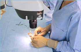
Hair transplant surgery has evolved significantly during its lifetime to become a fast, realistic and relatively stress-free process. Mantis inspection microscope has become the favoured tool of many practitioners.
Creating donor strips and graft dissection under magnification
 A modern hair transplant operation transfers around 1500 grafts, each containing between 1 and 4 hairs, to the treated area of the head. The grafts are taken from a donor strip removed from the back of the patient’s head.
The strip is typically between 9mm and 12mm wide and 75mm or more in length, depending on how many donor grafts are required. When the strip is removed, the resulting scar is closed and becomes imperceptible to all but the most detailed examination.
“The next phase of the operation requires great care and accuracy, and that’s where the Mantis stereo viewer comes in” according to Bill Eck of Physician’s Hair Institute in Tucson, Arizona.
The donor strip is slivered to create micrografts of 1 and 2 hairs, or minigrafts of 3 or 4 hairs.
“When cutting the donor strip into slivers in order to create the grafts for transplanting, we have to take care to isolate the exact number of hairs required for each graft. We must also avoid transecting the graft – that is, cutting through the root of the hair below the skin surface. Because we are working with individual hair follicles, this is difficult to accomplish with the naked eye”
, Eck explains.
Bill Eck is assistant to Dr. Sharon Keene, founder of the Physician’s Hair institute. After the donor strip of hair has been successfully removed, Dr. Keene will prepare the patient’s scalp to accept the grafts, while a team usually consisting of 2 technicians creates the range of micrografts and minigrafts specified during the primary consultation.
Preparing the scalp requires considerable surgical skill to achieve high quality of result. Away from the site of Dr. Keene’s work, the technicians painstakingly sliver the donor strip.
Once created, the grafts are placed in a petri dish, ready for the surgeon to transplant. In keeping with the way hair naturally grows, the patient’s front hairline is usually constructed using single-hair micrografts, while larger areas are covered with the minigrafts containing up to four hairs.
“Look at your own hair, or that of your colleagues,” suggests Eck. “You can identify single follicles at the hairline, but further back the follicles are more tightly grouped. We recreate these patterns in the transplanting process.”
Choosing the best equipment for hair transplant technicians
“Completing the treatment can take anything from 3 hours to over 10 hours, depending on factors such as the number of grafts required of the hair type,” continues Eck. “This can be quite fatiguing, especially when we are dealing with grey or ‘salt & pepper’ hair colours, for example. These are the most difficult to see and can appear transparent under the surgery lighting.”
He explains that to make these hairs more visible, and thus easier to process accurately, many techniques have been tried.
“We looked for new equipment that would help us do this better and a magnifying viewer was consistently the best solution. We now operate two Mantis instruments, supplied by Vision Engineering.”
TThe Physician’s Hair Institute surgery is equipped with 2 Mantis stereo inspection microscopes.
Mantis inspection microscope has become popular throughout the hair transplant industry, having been specified by several of the world’s top clinics, including the Laser Hair Institute in North Miami Beach, the Physician’s Hair Center in Clearwater, Florida and of course the Physician’s Hair Institute in Tucson.
The eyepieceless microscope provides a true stereo image with user selectable magnification from 2x to 10x, and can be used by operators wearing spectacles or contact lenses.
This combination of ergonomic design to permit optimal posture and optical performance to deliver high clarity images allow Mantis operators to work effectively and comfortably for extended periods – useful attributes for technicians separating follicular units during hair transplant surgery. Depth perception, illumination and colour rendition are important for easy, fast and accurate handling of grafts.
“Hands-free operation is, of course, vital”, says Eck. “Previously, we used magnifying glasses at 2x magnification, but Mantis gives a superior image and is far more comfortable to use.”
Although Vision Engineering offers Mantis microscope with a versatile selection of accessories to facilitate a range of applications, the base configuration has proved optimal for the Physician’s Hair Institute.
