The Institute of Stem Cells in the Treatment and Study of Monogenic Diseases (ISCMD), a Genopole® International Research Laboratory, in Evry near Paris, is utilising Vision Engineering’s Lynx stereo zoom microscope in their sealed extraction cabinets.
The eyepieceless optics of the Lynx means it can be placed behind the glass within the cabinets allowing operators to inspect the live stem cells in both brightfield & darkfield illumination up to 40x magnification.
ISCMD is based in Evry, near Paris in France. ISCMD is a research laboratory of Genopole® International and is dedicated to improving our knowledge and understanding of stem cells for the development of therapies for human diseases like Parkinson’s disease, Alzheimer’s disease, heart disease, stroke, arthritis, diabetes, burns and spinal cord damage.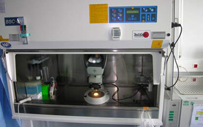
Minimising degeneration and contamination
During stem cell research, scientists need to implement a number of processes including the harvesting, inspection and the dissection of cells. To minimise degeneration and contamination, sealed extraction cabinets are used so that scientists can carry out their procedures from outside the cabinet, while the cells are magnified inside the cabinet by the Lynx biological microscope.
Traditionally, it would have been difficult to inspect the stem cells under magnification because the eyepieces of the microscope would have been inside the laminar flow cabinet.
However, as the Lynx does not have eyepieces but benefits from a viewer, the operator does not need to place their eyes directly in front of the binocular eyepieces.
Lynx Dynascope head
Vision Engineering’s patented Dynascope head is the key feature of the Lynx, allowing the system to deliver optimum clarity and pin sharp accuracy through a viewer instead of binocular eyepieces.
It is the ‘eyepiece-less’ viewer that allows the Lynx to be utilised behind the glass, where the operator can view the subject without the need to be directly in contact with the microscope.
Unlike conventional microscopes that have binocular eyepieces, the Lynx (with the ‘eyepiece-less’ viewer) allows operators to wear prescription spectacles, even while the operator is viewing the subject through the microscope, when it is inside the laminar flow cabinet.
Cell dissection under high magnification
The scientists at ISCMD concentrate on harvesting the stem cells so that they can be used for further research. Usually the stem cells are harvested within incubators but, once the cells have been successfully harvested, they require the scientists to dissect the samples to separate the cells using a micro manipulation pipette. This delicate process requires high magnification and superb clarity, to enable the scientists achieve accurate dissections.
In addition to the high clarity through the unique Dynascope® head, the Lynx research microscope enhances the contrast of the stem cells by the tilting mirror within the substage of the unit.
This creates a pseudo effect for both darkfield and brightfield maximising the contrast of the cells for both inspection and manipulation. Both the darkfield and the brightfiled are important features for both dissection and inspection.
Inspecting the stem cells for their structure, any granulation and the general morphology of the cells is important for maintaining the continuing high quality of the samples. If the cells had started to degenerate and the morphology of the cells were not up to the required standard, the time and money wasted using them for research would be costly.
