
Renishaw plc employs the use of Vision Engineering’s Mantis® stereo viewer to inspect dental frameworks for finished quality, precision and fit.
Renishaw has been manufacturing innovative measuring solutions for over 33 years and has recently identified the need within dentistry for a refined and accurate process for crown and bridge framework manufacture.
In doing this, Renishaw has designed and developed incise™ – a revolutionary dental CAD/CAM system that focuses on accuracy at every stage of the process, beginning with the first impression taken by the dentist.
Digital checking procedures
To support its own digital checking procedures, Renishaw uses Vision Engineering’s range of Mantis® stereo viewers to assist with all aspects of the process, from quality assessment of machine parts to visual inspection of the finished frameworks.
Renishaw’s Dental Products Division has taken a radical review of the conventional way dental restorations are manufactured. From this, the incise™ process was born, providing an innovative solution to the manufacture of all-ceramic parts.
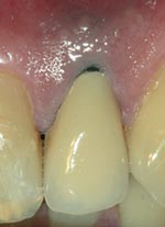
Figure 2.
Traditionally, the dental industry has used metal in the production of crown and bridge frameworks. Occasionally metal crowns can cause reactions resulting in gum recession and exposure of unattractive metal collars that are prone to decay collection and staining.
Renishaw’s incise™ process uses a strong, hard ceramic material called zirconia (see figure 2). The material is biocompatible so is extremely friendly to oral tissues encouraging better oral health. The metal-free core gives the benefit of a more natural, translucent appearance.
The accuracy of marginal fit that the incise™ process can achieve means increased resistance to decay and staining, resulting in a longer lasting restoration. To optimise the accuracy of the whole process from start to finish, Renishaw has been working closely with dentists and dental technicians to improve the overall quality of the finished restorations.
Renishaw provides a set of guidelines to help eliminate errors at each stage of the impression and die process, ensuring the precision of the finished reconstruction.
Dental restoration process overview
Initially, the patient has a consultation where the dentist provides a prescription for reconstruction to restore ideal jaw function or to improve aesthetics. The teeth are then prepared and, using Renishaw’s proven techniques and materials, the dentist takes impressions of the teeth and surrounding tissues.
The patient is fitted with temporary restorations to protect the prepared areas and the impressions are sent to the dental laboratory, together with any other relevant information on the patient’s clinical situation.
Even at this early stage, it is vital that the impressions are free of defects. From the impressions, stone models are made of the upper and lower jaws which are then articulated together to represent the patient’s bite.
The model of interest is sectioned and the prepared areas are removed, trimmed and measured using the incise™ scanning machine, which generates a three-dimensional representation of the stone model. The machine scans the model using a contact scanning probe, digitising the complex form of the preparation replica.
Data from the scan is then sent electronically to Renishaw’s milling centre in Gloucestershire where the zirconia framework is manufactured.
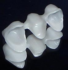
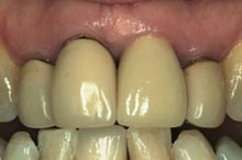
Figure 3.
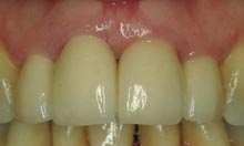
Figure 4
Renishaw incise™ frameworks are supplied with a certificate of conformance, showing an analysis of the framework fit to the original stone model. The zirconia framework and the certificate are packaged and sent back to the dental laboratory for porcelain build-up. The finished restoration is returned to the dentist along with the certificate.
The dentist will then complete the process by removing the temporary and fitting the incise restoration, checking the colour match and contacts with surrounding teeth and the essential marginal fit. When the dentist and patient are happy, the crown or bridge is permanently cemented in place.
Figure 3 shows four anterior porcelain jacket crowns, three of which were 30 years old. It is obvious where the poor fit of the crowns has caused the gums to recede, exposing dark margins & collecting decay.
Figure 4 shows the same patient with newly fitted incise™ crowns. The gums look extremely healthy and the aesthetic result is superb.
Before the ceramic frameworks are dispatched back to the laboratory, they are inspected using the Mantis range of stereo systems from Vision Engineering.
Renishaw has invested in a Mantis Elite stereo viewer and a number of Mantis Macro viewers with straight through viewing. After the parts have been machined, they undergo a final inspection. The cold bright white LED light of the Mantis range provides adequate illumination to inspect the uniformity of the parts.
How Renishaw got the perfect fit
When the project was in its infancy, Renishaw was using several methods of inspection including loupes. Although this method gave adequate magnification, the practicalities of inspecting through such equipment was not ideal, especially for long term use.
Bryan Austin, Director & General Manager of the Dental Products Division at Renishaw visited a number of dental laboratories during the early stages of the project and immediately identified how they were using the ergonomic Mantis stereo viewer from Vision Engineering. Bryan Austin explains:
“With the hours that are spent by production staff peering into eyepieces of microscopes or loupes, it made sense to invest in equipment that provided maximum comfort as well as optimum clarity.
“The Mantis delivered everything we needed to successfully carry out our visual inspection process. With the certificate of conformance that we are providing to our customers, it is imperative that we produce crowns and bridges that are a perfect fit.
“We use the Mantis to inspect the machined parts giving us total confidence that our incise™ scanning machine is producing accurate dimensions, resulting in prosthodontic parts that are a precise representation of the patient’s teeth”.
Bryan goes on to explain how having an inspection station changed the whole mindset of the inspector. “After we designed the inspection station using the Mantis, we found that when the inspectors physically took the framework over to the system and initiated the inspection procedure, it changed their whole psyche”.
The incise™ scanning system is validated to BS EN ISO10360 part 4 (an international standard relating to the accuracy of contact scanning systems) and all restorations are produced under a BS EN ISO13485 quality system.
With Renishaw introducing innovative measurement solutions and proven technology like the incise™ system into the medical devices industry, including dental technology, the benchmark has now been set for high precision and quality.
